Contact information: Registration: +48 41 367 4850
Main office: +48 41 367 4860
Fax: +48 41 367 4887
e-mail: zmnsco@onkol.kielce.pl
Head of the Department: Professor Janusz Braziewicz
Scientific advisor: Professor Leszek Królicki
The Department of Nuclear Medicine with Positron Emission Tomography (PET) Unit comprises:
-
Scintigraphy Laboratory,
-
PET/CT Laboratory,
-
Radiological Protection Laboratory
The Department of Nuclear Medicine with Positron Emission Tomography (PET) Unit as one of the few in Poland performs diagnostic tests using isotope techniques conducting synthesis of radiopharmaceuticals at its laboratory. We conduct scintigraphy and PET / CT examination, both structural and functional. The first scintigraphic examination was conducted in autumn 1992, using a dual detector gamma camera Multispect. Since then we have been systematically modernizing our diagnostic equipment. Annually, we conduct more than 3 thousand various types of scintigraphic studies, mostly single-photon emission computed tomography (SPECT). In 2008 the Department was equipped with a Biograph 64 PET / CT scanner. At present we carry out more than 3 thousand PET / CTs per year using 6 different radiopharmaceuticals. We are a subcontractor of the project implemented with the National Health Fund. We also examine patients referred by other medical facilities which cooperate with the NHF (the patient is not charged for the examination).
The Diagnostic Equipment of the Department includes:
PET / CT Laboratory:
-
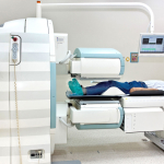
Biograph 64 PET/CT scanner,
-
Althea radiopharmaceuticals dispenser,
-
Automatic injector for CT with contrast agent,
-
Respiratory gating system,
-
Heart gating system,
-
MultiModality PET/CT diagnostic stations, Syngo Via server,
-
PACS data collecting system,
-
Radiopharmaceuticals Synthesis Laboratory,
-
Pharmaceuticals Quality Control Laboratory
Scintigraphy Laboratory:
-
SPECT/CT Symbia T2 camera,
-
Dual detector E-Cam camera,
-
Scintigraphic diagnostic stations,
-
Radiopharmaceuticals Synthesis Laboratory.
We perform the following PET/CT:
-
whole-body 18F-FDG PET/CT,
-
whole-body 18F-NaF PET/CT,
-
whole-body 18F-Choline PET/CT,
-
whole-body 18F-Dopamine PET/CT,
-
whole-body 18F-FET PET/CT,
-
whole-body 18F-FLT PET/CT,
-
whole-body Ga-68 DOTATE PET/CT,
Scintigraphic examinations:
1. Scintigraphy of the osteoarticular system:
-
Three-phase bone scans of a selected section of the skeleton,
-
SPECT scintigraphy,
-
Whole-body SPECT,
2. Cardiac muscle examination:
-
Myocardial perfusion (one or two-day examination using 99mTc-MIBI or 99mTc-tetrofosmin),
3. In vivo gated angiocardiography with 99mTc erythrocytes.
4. Lung scintigraphy:
-
99mTc angio-scintigraphy,
-
Planar perfusion lung scintigraphy (99mTc)
-
SPECT - Single Photon Emission Computed Tomography
5. Kidney examination:
-
Static planar kidney scintigraphy and Single Photon Emission Computed Tomography (99mTc-DMSA),
-
Filtration renal kidney scintigraphy (99mTc-DTPA),
-
Extraction renal kidney scintigraphy (99mTc-EC).
6. Parathyroid scintigraphy (planar and subtraction) (99mTc-MIBI and 99mTcO4).
7. Thyroid scintigraphy:
-
With 99mTcO4,
-
With 99mTc-MIBI,
-
Single Photon Emission Computed Tomography,
-
Whole-body examination after administering 2,5mCi 131J and whole-body examination after administering a therapeutic dose of 131J,
-
With iodine uptake of 131I activity 4MBq
8. Salivary glands scintigraphy
-
Angio-scintigraphy of salivary glands,
-
Functional scintigraphy,
-
Planar and tomographic SPECT of salivary glands.
9. Scintigraphy upper gastrointestinal tract:
-
esophageal function testing (with 99mTc-DTPA, 99mTc-sulphur colloid),
-
gastroesophageal reflux testing (with 99mTc-DTPA, 99mTc-sulphur colloid),
-
scintigraphy of the lower gastrointestinal tract - Meckel's diverticulum.
10. Scintigraphy of the liver and biliary tract:
-
function testing,
-
with in vivo labeled erythrocytes,
-
SPECT
11. Lymphoscintigraphy:
-
Dynamic and static (location of sentinel lymph node)
12. Whole-body scintigraphy:
-
With 99mTc-DMSA
-
With 99mTc-MIBI
-
With gallium chloride
-
With Otreoscan-111In and 131J-MIBG.
Research projects:
‘Information platform for the fusion of heart imaging studies’ is Poland's first research project carried out jointly by the Department of Nuclear Medicine of the John Paul II Specialist Hospital in Krakow (project leader) and the Department of Nuclear Medicine with PET Unit of the Holy Cross Cancer Center in Kielce (project member). The project focused on the development and implementation of a common platform in the so-called virtual cloud, enabling archiving, sharing and advanced data processing of imaging studies performed in different centers and using a variety of diagnostic imaging techniques (PET, CT, cardiac magnetic resonance imaging, single photon emission computed tomography, magnetic resonance spectroscopy). With this solution, physicians obtain maximum accuracy of the tests and may consult patients at a distance. The patient receives a more accurate diagnosis without the need to repeat imaging examinations performed in various centers, because physicians have access to all imaging tests of the patient placed and secured in the virtual cloud. The patient avoids duplication of research and a medical facility does not bear any additional related costs. The project is aimed at the patients suffering from heart diseases who underwent stem cell therapy for myocardial regeneration. The project was initiated in mid-2014 for cardiac patients and currently also focuses on patients with oncological diseases and neurological diseases. It was financed with funds from the National Centre for Research and Development and EU funds.
We cooperate with:
Jan Kochanowski University in Kielce,
Medical University in Warsaw,
John Paul II Specialist Hospital in Krakow
H. Niewodniczańska Institute of Nuclear Physics of the Polish Academy of Sciences in Krakow

